Day: May 22, 2023

  • Knee Pain and Popping: Causes & Treatment

    Knee Pain and Popping: Causes & Treatment

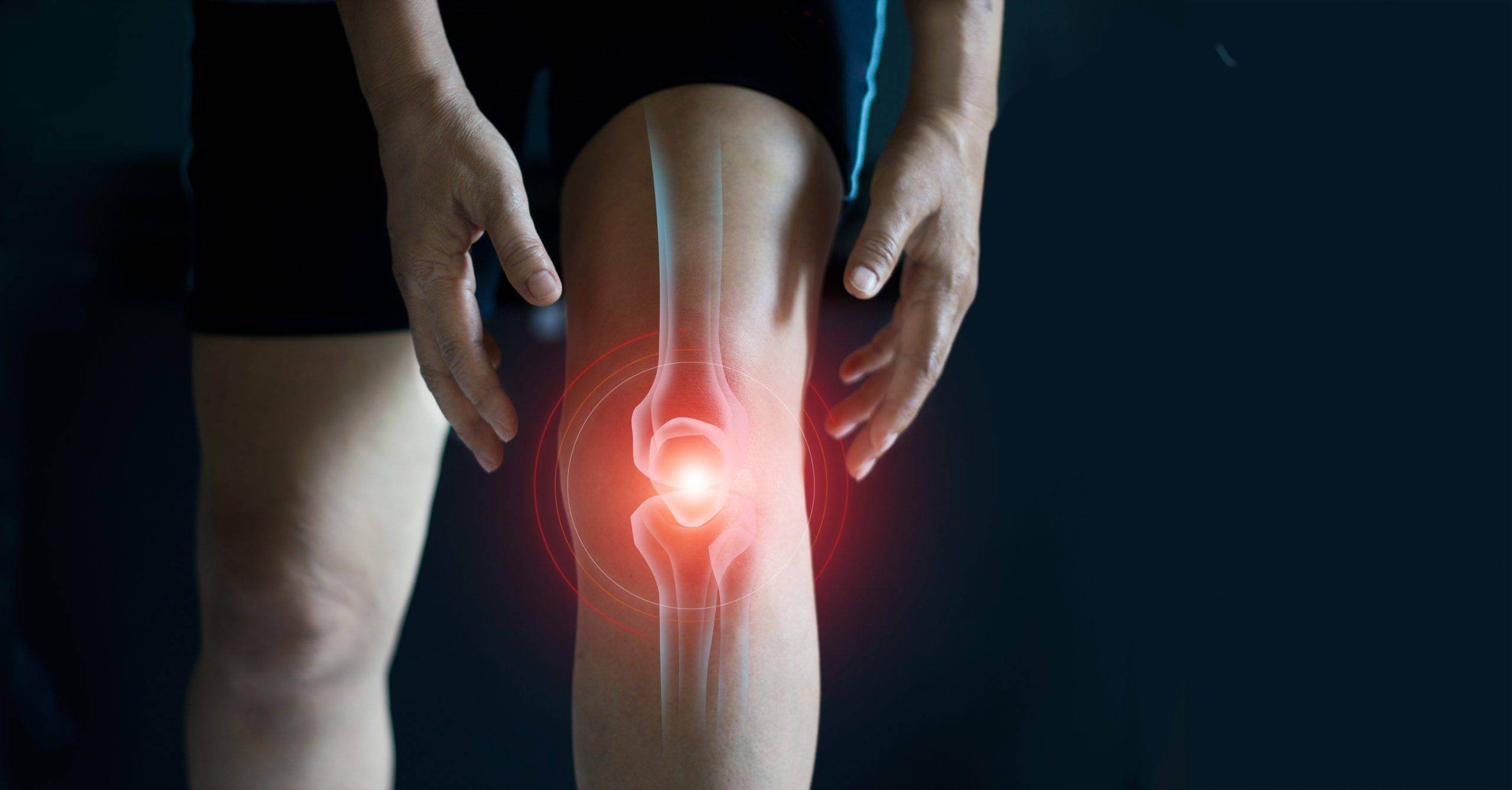
    A typical complaint that many people have at some point in their lives is knee discomfort and popping. Knee pain is defined as soreness or discomfort in or near the knee joint. It can range in severity and occasionally include popping sounds. When the knee is moved, popping may be present as a clicking, crackling, or snapping sound.

    The significance of comprehending the root causes and available remedies

    It’s crucial to comprehend the origins and available treatments for knee popping and pain for a number of reasons. First of all, knee discomfort and popping can have a serious negative influence on a person’s quality of life by restricting their capacity to accomplish everyday tasks, work, and engage in sports or other leisure activities. For symptoms to be reduced and function to be recovered, the underlying cause of knee pain must be found and treated efficiently.

    Second, distinct knee pain and popping causes call for various treatment modalities. Incorrect diagnosis or treatment can result in persistent pain, functional restrictions, and serious consequences. Therefore, having awareness of the potential causes and available treatments aids in enabling both patients and healthcare providers to make wise management strategy selections.

    Finally, being aware of the factors that contribute to disease and the available therapies enables people to take an active role in their own care. They can ask pertinent questions, participate in shared decision-making processes, and effectively express their symptoms to healthcare professionals by being aware of the options. This will improve the effectiveness of their therapy.

    How the Knee Works anatomically

    A succinct description of the knee joint’s architecture

    The thigh bone (femur), shinbone (tibia), and kneecap (patella) are all joined together at the knee joint, which is a complicated hinge joint. It is supported by a number of cartilage, muscle, ligament, and tendon structures. The following are the main parts of the knee joint:

    1. Femur: The thigh bone, which makes up the knee joint’s upper portion.
    2. Tibia: The bottom portion of the knee joint is made up of the shinbone.
    3. Patella: The patella, or kneecap, is a tiny bone that is a part of the patellar tendon and serves to guard the front of the knee joint.
    4. Articular Cartilage: The ends of the femur, tibia, and patella are covered in a smooth, protective cartilage called articular cartilage, which facilitates motion and lessens friction.
    5. Menisci: The knee joint’s medial and lateral menisci are two C-shaped sections of cartilage that serve as shock absorbers between the femur and tibia. They increase stability and evenly distribute stresses within the knee joint.

    Normal movement and function of the knee

    Walking, running, jumping, and many other actions are all made possible by the knee joint. The knee joint’s main motions include the following:

    1. Flexion: Knee flexion that brings the lower leg up towards the thigh.
    2. Extension: Straightening the knee, putting the lower leg back in place where it was before.
    3. Rotation: When the knee is flexed, the knee joint permits very minor internal and external rotation.

    For the knee joint to function properly, stability is essential. The knee joint’s surrounding ligaments, muscles, and tendons cooperate to provide stability and limit unnatural or excessive movement. By distributing stresses and absorbing shocks, the menisci and articular cartilage also help to maintain the joint’s stability and smooth motion.

    Therapy Alternatives

    Conservative Medical Measures

    1. Rest and Modification of Activities: Resting the knee and staying away from activities that make it pop or hurt will help lessen symptoms. It’s crucial to modify activities to put as little strain as possible on the knee joint to encourage healing.
    2. Pain management: NSAIDs, such as ibuprofen or naproxen, can help lessen pain and inflammation. Additionally, employing heat therapy or putting on ice packs helps relieve symptoms.
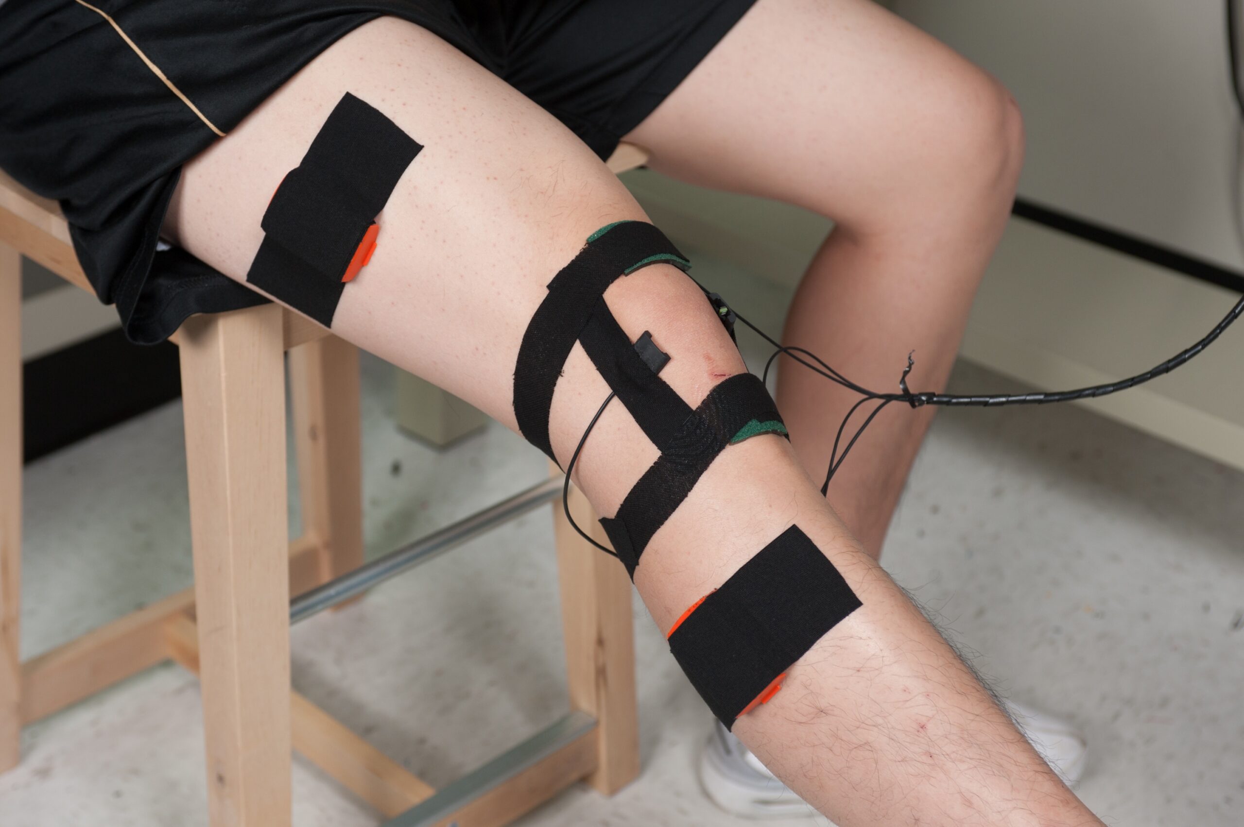
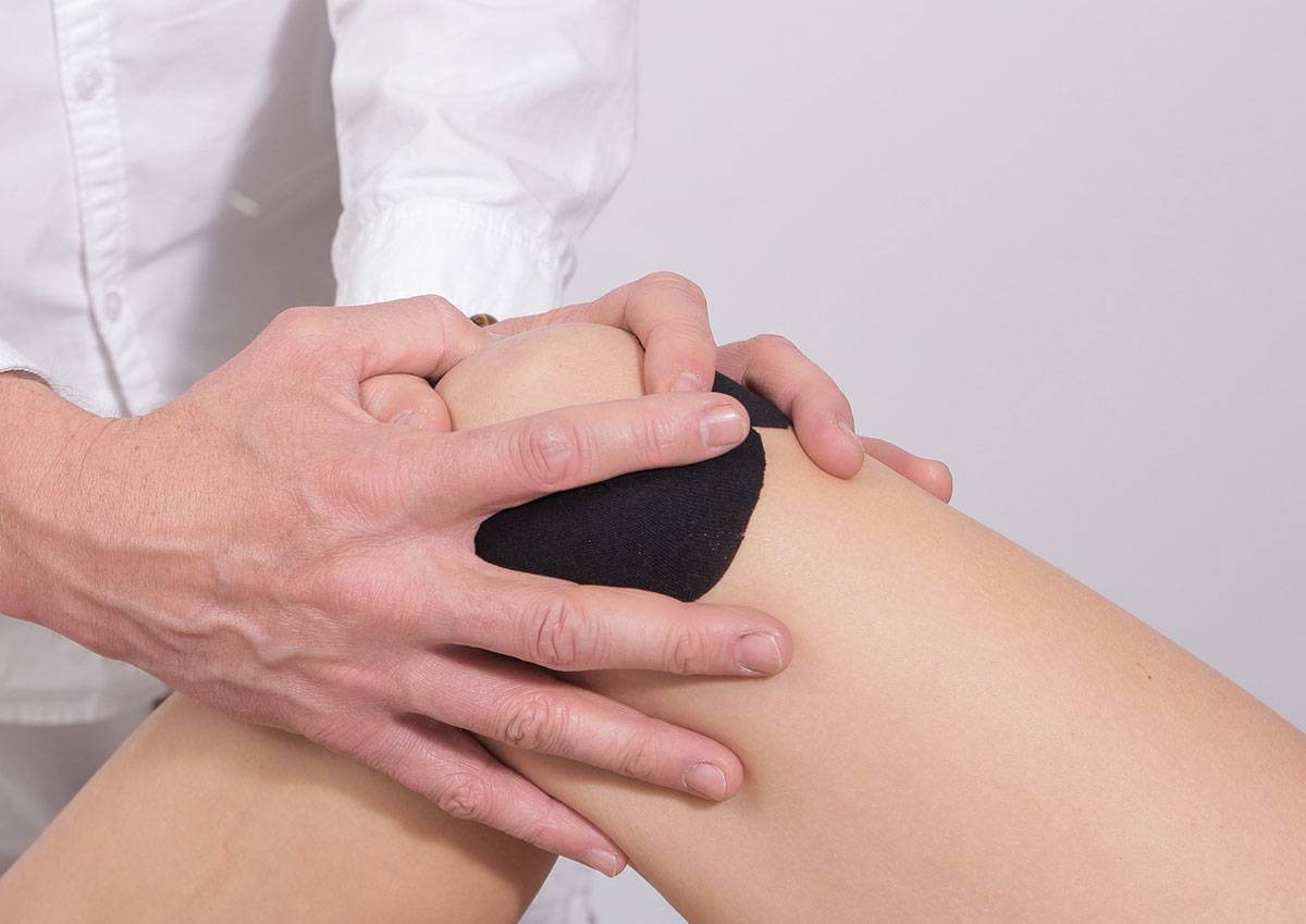
    3. Taping techniques: Bracing or taping techniques can offer support, stability, and pain reduction when used on the knee. These aids can ease symptoms and encourage appropriate alignment while performing tasks.

    Medications

    1. Analgesics for Pain Relief: Acetaminophen and other over-the-counter pain relievers can be used to treat mild to moderate knee discomfort. For more severe pain, prescription-strength painkillers may be advised.
    2. Anti-inflammatory Drugs: NSAIDs, such as ibuprofen or diclofenac, can lessen discomfort and inflammation related to diseases of the knee. Under medical supervision, these drugs should be taken.
    3. Injections: In some circumstances, injections for pain relief or inflammation reduction may be explored. Hyaluronic acid injections can lubricate the joint and help with osteoarthritis symptoms, while corticosteroid injections can temporarily relieve pain by lowering inflammation.

    Causes of Knee pain and popping

    1. Meniscal Tears: Meniscal tears are a frequent source of knee pain and popping, particularly in athletes or people engaged in occupations requiring quick turns or pivots. Between the femur and the tibia, the menisci can tear as a result of trauma or degeneration, producing discomfort, swelling, and a popping feeling.
    2. ACL, PCL, MCL, and LCL Ligamentous Injuries: The medial collateral ligament (MCL), lateral collateral ligament (LCL), anterior cruciate ligament (ACL), and posterior cruciate ligament (PCL) are some of the knee ligaments that can be injured. These injuries can result in knee instability, pain, and popping noises. Sports or other activities involving quick changes in direction or direct knee impact frequently result in these ailments.
    3. Bursitis: Around the knee joint, bursae—small sacs filled with fluid—cushion and lessen friction between tendons, muscles, and bones. Knee discomfort, swelling, and popping sensations can occur when these bursae become inflamed, which is often brought on by repetitive usage or direct damage.
    4. Plica Syndrome: The plica, a fold of synovial tissue in the knee joint, can become irritated or inflamed, resulting in plica syndrome. Particularly while engaging in repetitive knee bending or twisting movements, it might result in soreness, popping, or snapping feelings.
    5. Chondromalacia Patella: Also known as runner’s knee, chondromalacia patella is characterized by the deterioration and softness of the cartilage on the underside of the kneecap. It may be accompanied by popping or grinding sensations and cause knee pain, especially during tasks that require knee bending.

    Conclusion

    One’s quality of life can be greatly impacted by knee discomfort and popping, which can restrict mobility and obstruct daily tasks. For these symptoms to be effectively managed and relieved, it is essential to comprehend the reasons and available treatments. A medical expert must be consulted for a correct diagnosis and to choose the best course of action based on the patient’s unique needs. The discomfort can be reduced, knee function can be recovered, and general wellbeing can be enhanced with prompt and effective care.

    It’s important to keep in mind that this diagnosis chart for knee popping serves only as a general guide. To ensure a precise diagnosis and a customized course of treatment, always seek competent medical guidance.
  • Knee Pain Diagnosis Chart

    Knee Pain Diagnosis Chart

    Healthcare providers can analyze and diagnose the underlying causes of knee pain in patients using the knee pain diagnosis chart. It offers a well-organized framework for keeping track of crucial data, conducting physical exams, requesting pertinent diagnostic tests, and making the right diagnosis. The chart’s main goal is to assist medical professionals in comprehending the nature of the knee pain, spotting potential diseases or injuries, and determining the best course of therapy.

    The necessity of an accurate diagnosis for successful therapy

    A correct diagnosis is essential for knee pain treatment to be successful. The knee joint is a complicated structure, and a variety of ailments, from traumatic injuries to degenerative diseases and inflammatory disorders, can cause pain and discomfort. Without a clear diagnosis, treatment may be unsuccessful, resulting in the patient experiencing prolonged pain, functional restrictions, and a lower quality of life.

    Healthcare professionals can develop treatment plans to target the particular underlying cause of the knee pain by conducting a comprehensive and precise diagnosis. This could entail taking drugs, getting physical therapy, getting shots, getting aids, or having surgery. Furthermore, a precise diagnosis enables healthcare professionals to control patient expectations, deliver pertinent education, and give advice on self-care techniques like exercise and lifestyle changes.

    Visual Inspection

    Examination

    1. Swelling: Check for any felt or visible swelling near the knee joint, taking note of its location and degree.
    2. Deformities: Look for any obvious deformities in the knee joint, such as misalignment, bowing, or asymmetry.
    3. Changes in Skin: Examine the skin for any changes that might point to an injury or underlying ailment, such as pigmentation, redness, rashes, bruising, or lesions.

    Palpation

    1. Tenderness: Use a gentle touch to palpate the knee joint and its surroundings to locate any tender spots. Make note of their exact positions and any localised pain reactions.
    2. Warmth: Check the knee joint for increasing warmth or heat, which could signify infection or inflammation.
    3. Soreness along the joint line: Feel for soreness along the joint line of the knee to check for meniscal or ligament damage.

    Motion range

    By evaluating the patient’s capacity for flexion, extension, and rotational motion, determine the patient’s knee joint range of motion. If required, measure the movement’s degrees using a goniometer. Keep track of any restrictions, discomfort, or pain you had while undergoing the range of motion exam.

    Joint stability tests

    Assess the stability of the ligaments supporting the knee joint by performing stability tests. These examinations look for laxity or instability and can reveal ligamentous injuries. The anterior and posterior drawer tests, the valgus stress test, and the varus stress test are typical stability tests.

    Lab Examinations

    CBC (complete blood count)

    A basic blood test called a CBC can assist discover underlying disorders that may be the cause of knee discomfort while also providing information about the patient’s general health. Red blood cells, white blood cells, and platelets are just a few of the blood components that are measured. An inflammatory or infectious disease may be indicated by abnormalities in the complete blood count (CBC), such as an elevated white blood cell count or anemia symptoms.

    ESR, or the erythrocyte sedimentation rate.

    ESR is a blood test that gauges how quickly red blood cells accumulate in a tube over a predetermined period of time. It is a generalized indicator of inflammation and can assist reveal the presence of a bodily ailment that is causing inflammation. An increased ESR may be a sign of systemic inflammatory disorders such inflammatory arthritis.

    Protein C-reactive (CRP)

    Another blood indicator of inflammation is CRP. It is created by the liver in response to bodily inflammation. Elevated CRP levels can provide evidence for an inflammatory disorder causing knee discomfort by indicating the existence of acute or chronic inflammation.

    Analysis of joint fluid

    Synovial fluid from the knee joint is removed and subjected to joint fluid analysis, sometimes referred to as arthrocentesis. It is carried out to assess the properties and make valuable diagnostic data about the fluid. As part of the examination, the fluid is checked for symptoms of infection, crystals (which can be a sign of illnesses like gout), and cell count (which might be a sign of inflammation or infection). Additionally, it can be used to identify various forms of arthritis or other problems involving the joints.

    Prognosis

    Possible results

    The underlying reason, the seriousness of the ailment or damage, and individual characteristics all affect the prognosis for knee discomfort. Possible results include:

    1. Full recovery: In some circumstances, patients who receive the proper care and therapy can completely eradicate their knee pain and resume their regular activities without any restrictions.
    2. Partial recovery: Some people may have some symptom reduction, increased function, and pain relief, but they may continue feel minor discomfort or have some activity restrictions.
    3. Chronic condition treatment: To reduce symptoms and preserve function, certain knee disorders, such as osteoarthritis or chronic tendinopathy, may call for long-term management techniques. Even while a full cure might not be achievable, good management can help people live better lives and experience less pain.

    Recovery period

    Depending on the underlying cause and the patient’s response to treatment, the time it takes for knee pain to go away might vary greatly. It can take anywhere between a few weeks for small wounds and several months for more serious ailments or surgery. The patient’s age, general health, adherence to treatment regimens, and participation in rehabilitation programs are all factors that affect how quickly they recover.

    Conclusion

    The knee pain diagnosis chart offers a thorough and organized method for assessing and diagnosing patient knee discomfort. Healthcare practitioners can make an accurate diagnosis by compiling patient data, performing a comprehensive physical examination, ordering pertinent diagnostic tests, and taking into account differential diagnoses.

  • Knee Pain Diagnosis, Symptoms & Treatment

    Knee Pain Diagnosis, Symptoms & Treatment

    Knee discomfort can have a substantial negative influence on a person’s quality of life by restricting mobility and interfering with daily activities. To give the right care and avoid further issues, it is critical to precisely evaluate the underlying cause of knee discomfort. Early detection and intervention can reduce discomfort, enhance function, and shield the knee joint from long-term harm.

    Overview of typical knee pain causes:

    The complex knee joint bears a great deal of weight and is subjected to repeated motions. Knee discomfort can result from a number of sources, such as:

    1. Osteoarthritis: This ailment affects the cartilage that cushions the bones in the knee joint, and it is a degenerative joint disorder. Pain, stiffness, and edoema may result from it, which frequently affects elderly people.
    2. Rheumatoid arthritis: It is an autoimmune condition that affects the joints, especially the knee joint, and produces inflammation. Chronic discomfort, swelliness, and joint deformity can all be side effects of rheumatoid arthritis.
    3. Ligament injuries: ACL, MCL, PCL, and LCL are the four primary ligaments that stabilize the knee joint. Ligament injuries. Knee discomfort, instability, and mobility issues may come from accidents or sports-related injuries to these ligaments.
    4. Meniscus tears: In the knee, the meniscus, a C-shaped portion of cartilage that acts as a shock absorber, tears. Sudden twisting or turning movements can result in meniscus tears, which can hurt, swell, and make it difficult to bend or straighten the knee.
    5. Patellofemoral pain syndrome: Also referred to as “runner’s knee,” this condition causes discomfort in the region in front of the knee, particularly behind or around the kneecap. Anyone who engages in repetitive knee motion activities, such as athletes, is usually seen with it.

    In order to get the proper diagnosis and create a successful treatment strategy, it is essential to understand the common causes of knee pain. It is always advised to speak with a medical expert for a correct assessment and diagnosis of knee discomfort.

    Signs of Knee Pain

    Pain and discomfort

    Feeling pain or discomfort in or around the knee joint is one of the main signs of knee pain. Aching, sharp, or dull throbbing sensations may also accompany the pain, which ranges in intensity from mild to severe. Walking, running, or bending the knee might make the pain worse. The discomfort could be intermittent or ongoing.

    Swelling and inflammation

    Swelling and inflammation are frequent components of knee discomfort. The affected knee may feel sensitive to the touch, be clearly swollen, and have elevated temperature. The buildup of fluid in the joint causes swelling, which is a sign of an underlying problem or injury. Weakness or instability: The knee that is experiencing discomfort may feel weak or unstable. People may feel as though their knee is “giving way” or that they cannot sustain their weight adequately. When there is instability, it can be difficult to maintain balance and to perform tasks that call for stability, such playing sports or walking on uneven terrain.

    Redness and warmth:

    The skin around the affected area may appear red and feel warm to the touch due to inflammation brought on by knee discomfort. The redness and warmth felt are a result of increased blood flow to the area brought on by the inflammation.

    Diagnostic Techniques

    Medical history and physical exam

    Gathering comprehensive medical information and performing a complete physical examination are required as the first steps in evaluating knee discomfort. The medical expert will inquire about the start and persistence of symptoms, prior injuries, pain-inducing or -alleviating activities, and any pertinent medical problems. A physical examination will also be done, looking for any indications of swelling, discomfort, range of motion, stability, and muscle strength in the knee. This aids in locating plausible reasons and limiting the range of possible diagnoses.

    Imaging tests (X-rays, MRI, CT scan)

    Imaging tests, such as MRI and CT scans, are frequently used to provide fine-grained images of the knee joint, assisting in the diagnosis and assessment of knee discomfort. These tests could involve:

    1. X-rays: X-rays show a two-dimensional image of the knee’s bones. They can aid in spotting fractures, anomalies in the bone, osteoarthritis of the joints, or indications of infection.
    2. Magnetic Resonance Imaging (MRI): The structures within the knee joint, including the bones, cartilage, tendons, ligaments, and soft tissues, can be visualized in great detail using magnetic resonance imaging (MRI), which employs strong magnets and radio waves. Ligament rips, meniscus injuries, cartilage degradation, and other anomalies of soft tissue can all be found with MRI.
    3. Computed Tomography (CT) scan: A cross-sectional image of the knee joint is produced by a CT scan by combining a number of X-ray images collected at various angles. CT scans offer comprehensive details about the bones, joints, and encircling structures. When evaluating complex fractures, bone anomalies, or when additional clarity is required for surgical planning, they are especially helpful.

    Joint aspiration

    Also referred to as arthrocentesis, joint aspiration is taking a tiny sample of synovial fluid from the knee joint for testing. Diagnoses for illnesses including infection, gout, or inflammatory arthritis are made with the aid of this method. For indications of inflammation, infection, crystals, or other anomalies, the fluid sample is evaluated.

    Therapy Alternatives

    Moderate strategies

    1. Rest and altered activity patterns: Resting the knee and avoiding pain-inducing activities can aid in the healing process. It is possible to lessen pain and prevent additional injury by cutting back on or altering activities that place an excessive amount of strain on the knee joint.
    • Exercises and physical therapy: Physical therapy is essential in the management of knee discomfort. To bolster knee stability overall, increase flexibility, and strengthen the muscles around the knee, a physical therapist can create a program of exercises specifically for you. Exercises can be low-impact aerobic activities, strengthening exercises, and range-of-motion exercises.
    • Pain management: Pain management methods can help with knee pain. They might consist of: medicines: No steroidal anti-inflammatory medicines (NSAIDs), such as ibuprofen or naproxen, can aid with pain relief and inflammation reduction. A medical expert might occasionally advise taking prescription drugs. Ice/heat therapy: Ice packs can help relieve discomfort and swelling in the affected area. Warm baths or compresses, which are forms of heat therapy, can promote blood flow and reduce stiffness.

    Conclusion

    In conclusion, knee discomfort can have a substantial influence on a person’s quality of life, thus it’s critical to accurately identify and treat the underlying reasons. People can seek the right medical care, take action to relieve pain, and improve knee function by being aware of the significance of diagnosing and treating knee pain.

  • The Role of Posture in Preventing Lower Back Pain

    The Role of Posture in Preventing Lower Back Pain

    The term” lower reverse pain” describes any discomfort or pain that’s felt in the lumbar area, which is the lower part of the backbone that’s  positioned between the  caricature  pen and the  bottom of the pelvis. The inflexibility may vary, varying from a patient  pang to a crushing  torture. Acute lower reverse pain might continue for a many days to a couple of weeks, or it can be  endless and endure for three months or longer.

    Cause of back pain:

     Having lower back discomfort can significantly affect a person’s everyday life and general well- being. It can circumscribe movement and make it delicate to carry out diurnal tasks like bending over, lifting, or walking. The discomfort may hamper productivity at work, involvement in rest hobbies, and indeed sleep quality. Reduced quality of life, internal discomfort, and frustration can all affect from lower reverse pain.

    Management and Self-Care

    Operation and tone- Care When deciding whether to seek medical attention, it’s vital to take into account the strictness of the lower rear pain and how it responds to tone- care ways. In general,  Acute pain Medical intervention may not be  demanded right down if the pain is minor to moderate and gets better with tone- care styles in a  numerous days to a couple of weeks.  habitual or worsening pain It’s advised to seek medical attention if the pain lasts further than three months or gets worse despite  tone- care measures.  Pain that is violent or has a fast onset or that is accompanied by other unsettling symptoms should be treated right formerly if they arise as a result of trauma or an injury.

     When to consider hunt medical attention after product some attempts at tone- defence.  Indeed, if original  tone- care procedures have been proved, there are some situations in which medical examination should be taken into an account. However,  impassiveness, Chinking, If lower  reverse pain is accompanied with radial pain.

     Implicit Serious Causes and Red Flags :

     Symptoms:

     A description of advising signs that could be major underpinning causes of rear pain  Red flags area unit advising signs and symptoms that could point to angstrom unit  dangerous  beginning condition  trouble lower back pain.   unforeseen onset of excruciating pain with no egregious cause. Fever and feverish pain or unanticipated weight loss Symptoms  Certain lower  reverse pain symptoms bear  exigency medical intervention. These correspond of a lack of bladder or bowel control chronic or recreating lower reverse pain habitual or intermittent Lower Back Pain. How long- term health is compact by prevailing lower  reverse pain  Habitual lower rear pain is defined as patient or constant lower rear pain that lasts for at least three months. The internal and physical well- being of that existent could be seriously affected. habitual pain can lead to lower movement, dropped morals of life, and restrictions on  quotidian  exertion. likewise, it might worsen internal health problems including guilt and anxiety.  Also,  habitual lower  reverse uncomfortableness could affect in functional confinement, difficulty falling asleep, and a general impairment in  fleshly fitness.

    When should someone with habitual or intermittent lower reverse discomfort seek medical attention? 

    It’s advised to seek medical attention if your lower aft discomfort is habitual or intermittent. Some signs that take medical trial view longer than three months of discomfort Significant effect on everyday tasks, employment, or quality of life. In order to estimate and treat habitual lower reverse pain, a multidisciplinary approach is generally used. Typical choices can include. Physical examination and evaluation of medical history. To determine implicit causes and contributing factors, the healthcare expert will estimate your symptoms, carry out a physical test, and ask about your medical history.

    Need to visit to the doctor:

    It’s important to understand when to get lower back pain treatment for a number of reasons. In the  morning, it enables accurate assessment and  opinion of the underpinning source of the pain. Some lower  reverse pain causes, like spinal cord  contraction or infections, demand  critical medical care. Second, prompt opinion and treatment can prop  in reducing discomfort, avoiding complications, and  speeding  the  mending process. Last but not least, getting medical attention gives you the chance to get advice on how to manage your pain, change your life, and take preventives to lessen the liability of  intermittent attacks.

    Severe pain:

    If you experience violent, excruciating pain that does not subside with rest or unpleasant pain details, it is advisable to seek medical help.

    Trauma or injury:

    If your back pain is the result of a fall, accident, or other significant injury, it’s important to seek medical attention to rule out fractures, sprains, or other serious conditions.

    Numbness or weakness:

    If you have numbness, tingling, or weakness in your legs, this could be a sign of contraction or damage like a herniated disc. These symptoms bear diagnosis by a health care professional.

    Loss of bladder or bowel control:

    If you experience difficulty controlling your bladder or bowel movements along with rectal pain, this may indicate a condition called cauda equina pattern, which requires immediate medical attention.

    Developing or worsening symptoms:

    If your back pain is worsening, or if it is associated with other associated symptoms such as weight loss, fever, or night sweats, it is important to see a doctor.

    History of cancer or osteoporosis:

    If you have a history of cancer or osteoporosis and you witness new-onset back pain, it’s important to get evaluated right away because it could be related to a serious condition. Prolonged Duration If your back pain persists for more than several weeks despite conservative treatment, it is advisable to consult a health care professional for further evaluation and guidance.

    Important points:

    Flash back, developing and maintaining good posture is an ongoing practice. It may take time to correct poor habits, but with  harmonious  trouble, you can gradationally ameliorate your posture and reduce the  threat of lower aftpain.bHowever, it’s  judicious to consult with a healthcare professional for a proper evaluation and guidance, If you  witness  patient or severe  reverse pain.

  • The Anatomy of Back Pain: What You Need To Know

    The Anatomy of Back Pain: What You Need To Know

    Back pain is a current health concern affecting millions of people worldwide. Understanding the deconstruction of reverse pain is pivotal for both cases and healthcare professionals to effectively manage and  help its  circumstance. In this composition, we will claw into the intricate details of  reverse pain, exploring its causes, common types,  threat factors, and available treatments. It can range from mild discomfort to debilitating pain, and understanding its anatomy is crucial for effective management and prevention. In this article, we will explore the various components of the back, common causes of back pain, and provide tips for alleviating and preventing this condition.

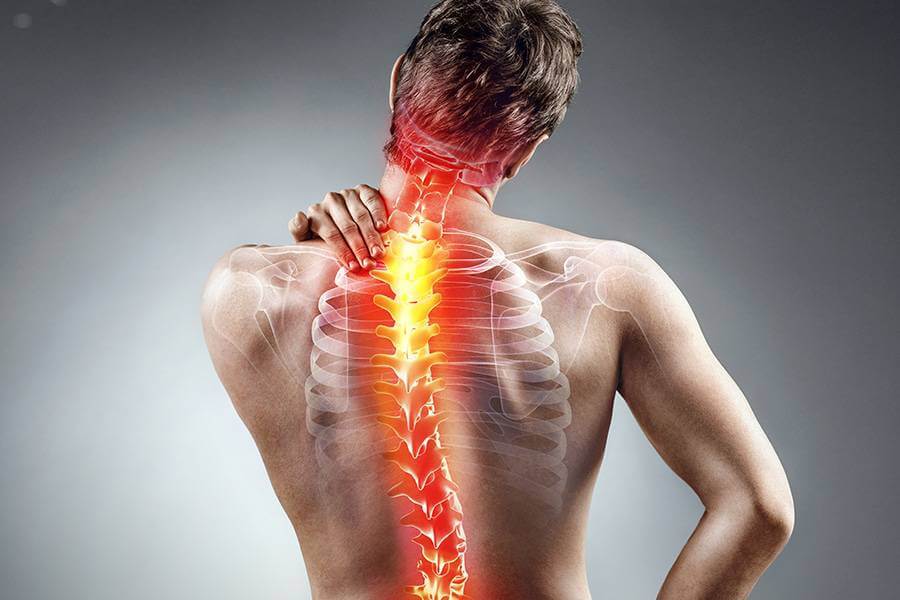
    Back pain is a common ailment that affects a significant portion of the population at some point in their lives. It can vary in intensity, duration, and location, often causing discomfort and limiting daily activities. To understand the anatomy of back pain, it is crucial to explore the structures involved and their potential sources of pain.

    Structure:

    The back is a complex structure composed of bones, muscles, ligaments, tendons, discs, and nerves. The spinal column, also known as the vertebral column, is a key component of the back. It consists of individual vertebrae stacked on top of each other, forming the spinal canal, which protects the spinal cord. There are four main regions of the vertebral column: cervical (neck), thoracic (upper back), lumbar (lower back), and sacral (pelvic region).

    The structure of the Back :

    The reverse is a complex structure composed of bones, muscles, ligaments, tendons, and discs. It consists of three main regions the cervical n( neck) region, thoracic(mid-back) region, and lumbar( lower  reverse) region. Each section has its own unique characteristics and functions.

    Cervical Region:

    The cervical  chine consists of seven chines labeled C1 to C7. It supports the head and allows for its mobility. The neck muscles and ligaments  give stability and  grease movement.

    Causes of Back Pain:

    Muscle strain or sprain

    One of the primary sources of back pain is muscle strain or sprain. The muscles in the back provide support and stability to the spine. When they are subjected to excessive stress, such as improper lifting, sudden movements, or poor posture, they can become strained or sprained, leading to pain and discomfort.

    Intervertebral disc problems

     Disc degeneration, herniation, or bulging can occur due to aging, injury, or repetitive stress, resulting in pressure on nearby nerves and causing pain.

    Spinal stenosis

    Spinal stenosis is another condition that can contribute to back pain. It is characterized by the narrowing of the spinal canal, which can compress the spinal cord and nerve roots. This narrowing may occur due to aging, arthritis, or other degenerative conditions. Individuals with spinal stenosis may experience back pain, numbness, tingling, and weakness in the affected areas.

    Structural abnormalities

    Structural abnormalities, such as scoliosis (abnormal curvature of the spine), kyphosis (excessive forward curvature of the upper back), or lordosis (increased inward curvature of the lower back), can also cause back pain. These conditions may be congenital or develop over time due to poor posture, muscle imbalances, or underlying diseases.

    Inflammation or irritation of the facet joints

    Additionally, back pain can arise from inflammation or irritation of the facet joints, which are small joints located at the back of the spine that allow for movement between adjacent vertebrae. Facet joint arthritis, injury, or mechanical stress can lead to pain in the affected area.

    The sacroiliac joint, which connects the sacrum to the pelvis, can also be a source of back pain. Dysfunction or inflammation of this joint can result in localized pain in the lower back and buttocks.

    Nerve impingement or compression

    Furthermore, nerve impingement or compression can cause radiating pain down the legs. Conditions such as herniated discs, spinal stenosis, or sciatica (irritation of the sciatic nerve) can lead to this type of pain, commonly referred to as radiculopathy.

    It’s worth noting that psychological factors, such as stress, anxiety, and depression, can influence the perception and experience of back pain. Emotional and mental well-being play a role in how an individual perceives and copes with pain.

    Ankylosing Spondylitis

    Inflammatory conditions can also contribute to back pain. Conditions like ankylosing spondylitis, which is a type of arthritis that primarily affects the spine, can cause chronic inflammation, stiffness, and pain in the back. Other inflammatory diseases like rheumatoid arthritis or infections of the spine can also lead to back pain.

    Muscle Imbalances

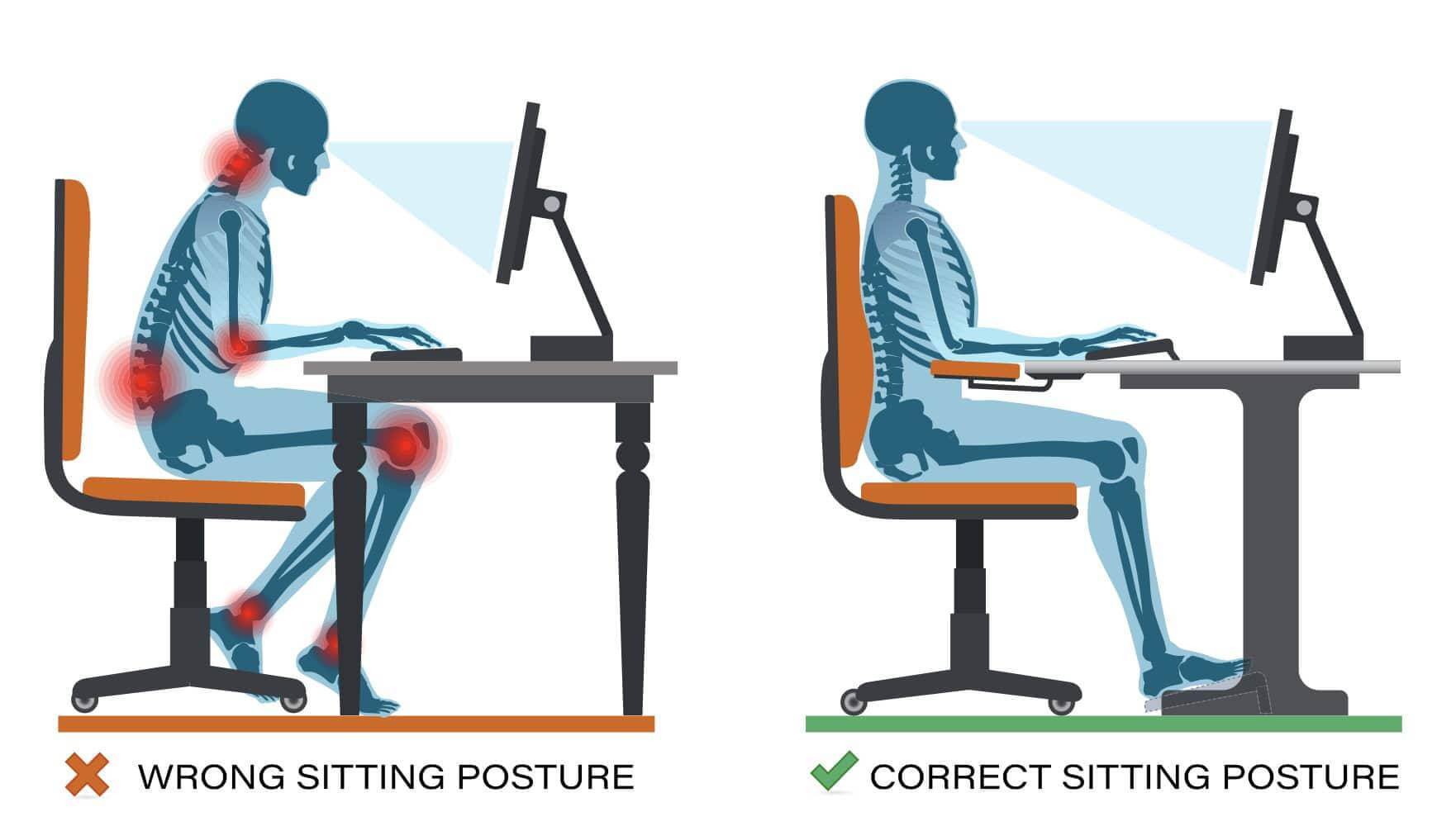
    Muscle imbalances and poor posture can play a significant role in the development of back pain. Weak core muscles, tight hip flexors, and imbalances between the muscles that support the spine can put excessive stress on the back, leading to pain. Poor posture, especially from prolonged sitting or standing in improper positions, can also strain the back muscles and contribute to pain.

    Psychosocial factors

    Psychosocial factors can significantly influence the experience of back pain. Stress, anxiety, depression, and other emotional factors can amplify the perception of pain and contribute to its chronicity. Additionally, negative emotions and psychological distress can lead to muscle tension, poor coping mechanisms, and decreased physical activity, all of which can worsen back pain.

    Lifestyle factors

    Certain lifestyle factors can increase the risk of developing back pain. Sedentary behavior, lack of regular exercise, and excessive weight can strain the back and contribute to pain. Smoking, which can impair blood flow and reduce the body’s ability to heal, has also been linked to an increased risk of back pain and slower recovery.

    Occupational factors

    Occupational factors can play a role in the development of back pain. Jobs that involve heavy lifting, repetitive movements, prolonged sitting or standing, or awkward postures can put excessive strain on the back and increase the risk of developing pain. Poor ergonomics, such as improper workstation setup, can also contribute to back pain in the workplace.

    Diagnosis

    Diagnosing the specific cause of back pain often requires a comprehensive medical evaluation, which may include a physical examination, medical history review, imaging tests (e.g., X-rays, MRI scans), and sometimes, specialized diagnostic procedures.

    Treatment

     It can involve conservative measures such as rest, physical therapy, pain medications, and lifestyle modifications. In some cases, more invasive interventions like injections, minimally invasive procedures, or surgery may be necessary.

    Conclusion

    In conclusion, back pain is a multifaceted condition that can arise from various structures in the back, including muscles, discs, joints, and nerves. Understanding the anatomy of back pain is crucial for proper diagnosis and management. If you experience persistent or severe back pain, it is recommended to consult a healthcare professional for an accurate evaluation and tailored treatment plan.

  • Top 10 Exercises to strengthen your Back and reduce Pain

    Top 10 Exercises to strengthen your Back and reduce Pain

     Then are a many general tips that may help palliate back pain.

    • Rest and avoid conditioning that worsen the pain It’s generally a good idea to take a break from conditioning that aggravate your reverse pain and allow your body to heal. 
    • Apply heat or cold wave remedy Applying a heating pad or a cold pack to the affected area may help reduce pain and inflammation. You can experiment with both options to see which works stylish for you.
    • Maintain good posture. Exercise and stretch Gentle exercises and stretches can help strengthen the muscles in your reverse and ameliorate inflexibility. still, it’s important to consult with a healthcare professional or a physical therapist to determine which exercises are suitable for your specific condition.
    • Over-the-counter pain relievers Nonsteroidalanti-inflammatory medicines ( NSAIDs)  similar as ibuprofen or acetaminophen can help  palliate pain and reduce inflammation. still, it’s stylish to consult with a druggist or your croaker before taking any drug to  insure it’s safe for you. 
    •  Use proper lifting ways When lifting heavy objects, flash back to bend your knees and lift with your legs rather than your reverse. Avoid wringing movements while lifting.   Maintain a healthy weight redundant weight can put fresh strain on your back. However, losing weight through a combination of a healthy diet and regular exercise may help reduce back pain, If you are  fat.   Flash back, these suggestions are general advice, and it’s pivotal to consult with a healthcare professional to determine the underpinning cause of your reverse pain and admit applicable treatment.

    Causes; Symptoms and Treatment:

     Back pain refers to discomfort or pain felt in the area of the reverse, which is located between the neck and the buttocks. It’s a current condition that can range from mild to severe and can be acute or habitual in nature. 

    Causes:

    1. There are causes of reverse pain, including muscle strains or sprains, poor posture, herniated discs, spinal stenosis, osteoarthritis, osteoporosis, cadaverous irregularities (similar as scoliosis), and traumatic injuries. also, life factors like sedentary geste

              rotundity, and lack of exercise can contribute to the development of reverse pain. 

    Symptoms:

    •  The symptoms of reverse pain can vary extensively and may include localized or radiating pain, stiffness, muscle spasms, reduced inflexibility, and difficulty in performing diurnal conditioning. In some cases, back pain can also be accompanied by impassiveness, chinking sensations, or weakness in the legs.
    •    Treatment for reverse pain depends on the underpinning cause and inflexibility of the condition. It may include tone- care measures similar as rest, hot or cold remedy, gentle stretching exercises, and untoward pain specifics. In more severe cases or when conservative measures fail, medical intervention like physical remedy, tradition specifics, injections, or, infrequently, surgery may be recommended.   Prevention of reverse pain involves maintaining a healthy weight, rehearsing good posture, engaging in regular physical exertion, using proper lifting ways, and avoiding prolonged sitting or standing in one position. It’s also important to address any ergonomic issues in the plant or at home that may contribute to reverse pain.   still, it’s judicious to consult with a healthcare professional for an accurate  opinion and applicable  operation, If you are  passing  patient or severe  reverse pain.

    Need to visit to the docter:

    It’s important to understand when to get lower back pain treatment for a number of reasons. In the morning, it enables accurate assessment and  opinion of the underpinning source of the pain. Some lower reverse pain causes, like spinal cord  contraction or infections, demand  critical medical care. Second, prompt  opinion and treatment can  prop  in reducing discomfort, avoiding complications, and  speeding  the  mending process. Last but not least, getting medical attention gives you the chance to get advice on how to manage your pain, change your life, and take  preventives to lessen the liability of  intermittent attacks.

    Top 10 Exercises to strengthen your Back and Reduce Pain:

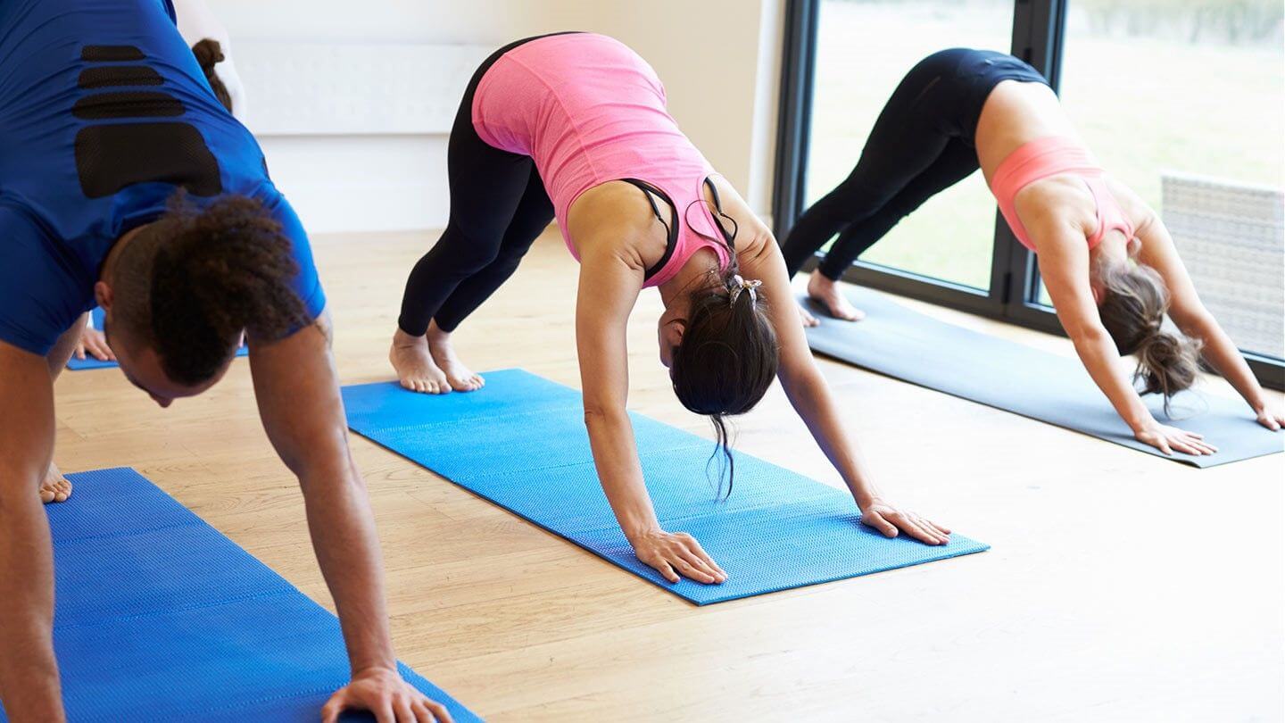
    Strengthening the back muscles is important to reduce pain and maintain a healthy chin. The following are ten exercises that can help you relieve your inversions and pain. Return to consult a health care professional before starting any exercise program, especially if you have a pre-existing condition or injury.

     There are some Top Exercises to Strengthen Your Back and Reduce Pain:

    Deadlifts:

     Deadlifts target multiple muscles in your reverse, including the erector spinae, lats, and traps. Start with light weights and concentrate on maintaining proper form throughout the exercise.

    Bent-Over Rows:

     This exercise primarily targets the muscles in your upper reverse, including the rhomboids and lats. Use a barbell or dumbbells, depend forward at the hips, and row the weights towards your casket while keeping your reverse straight.

    Superman Pose:

     Lie facedown on the bottom with your arms extended in front of you. Lift your arms, casket, and legs off the ground contemporaneously, creating a” flying” position. Hold for a many seconds and also lower back down. This exercise targets the muscles in your lower reverse. 

    Bird Dog:

     Get along with all fours with your hands directly under your shoulders and knees under your stays. Extend your right arm forward while contemporaneously extending your left leg backward. Keep your core meshed and your reverse perpendicular. Return to the starting position and replacement part This exercise engages the core and stabilizes the reverse. 

    Bridge Pose:

     Prevarication on your backward with your knees fallacious and bases flat on the  bottom. Force through your bottom, engage your glutes, and lifts your hips off the ground until your body forms a straight line from your shoulders to your knees. Hold for a many seconds, also lower back down. Bridge pose strengthens the glutes and lower reverse muscles.

    Lat Pulldowns:

    This exercise targets the muscles in your upper reverse and helps ameliorate posture. Use a lat pulldown machine or resistance bands to perform the movement. Sit with your hands gripping the bar or bands above, also pull it down towards your  casket while keeping your  reverse straight. 

    Rear Plank:

    Sit on the bottom with your legs extended in front of you and your hands resting on the ground behind your hips, fritters pointing towards your bases. Lift your hips off the terra firma, creating a direct line from your head to your heels. Hold for a many seconds and also lower back down. The rear plank strengthens your entire reverse and core.  

    Wall Slides:

    Stand with you reverse against a wall and your bases hip- range piecemeal. sluggishly slide down the wall, bending your knees and keeping your reverse flat against the wall. Hold for a many seconds and also slide back over. This exercise strengthens the muscles in your upper reverse and improves posture. 

    Plank :

    Get into a drive-up position with your forearms on the ground, elbows directly under your shoulders. Absorb your core and keep your body in a direct line from head to toe. Hold this view for as long as you can while hold over proper form. The plank exercise targets the core, including the muscles that support the lower reverse. 

    Cat- Cow Stretch:

     Get down on all fours with your hands straight under your shoulders and knees under your hips. Arch your back over towards the ceiling,  put away your chin towards your  casket( cat  disguise). also, let your belly Gomorrah towards the bottom, lifting your head and tailbone( cow  disguise). Alternate between cat and cow acts for several reiterations. This stretch helps to rally and stretch the muscles in your reverse. 

    Important information about Exercises :

     Flash back to start with lighter weights or shorter holds if you are a freshman, and gradationally increase the intensity or duration as your reverse becomes stronger. It’s essential to hear to your body and stop any exercise that causes pain or discomfort.